Foot skeleton with ligaments and muscles
Foot skeleton with ligaments and muscles
Foot skeleton with ligaments and muscles
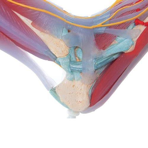
This anatomically detailed model of the foot and lower leg can be separated into 6 removable parts for detailed examination of the foot and ankle.
The foot skeleton has not only the bones, but also muscles, tendons, ligaments, nerves and arteries. The front view of the foot model has the extensor muscles of the lower leg. The tendons can be followed as they pass under the transverse and crucial crural ligaments all the way to their insertion points. In addition, all tendon sheaths in the foot area are visible. On the dorsal part of the foot, the gastrocnemius muscle is removable to reveal deeper anatomical elements. The sole of the foot is represented in three layers; the first layer showing the flexor digitorum brevis. This muscle can be removed from the foot revealing the quadratus plantae, the tendon of the flexor digitorum longus and the flexor hallucis muscle. This second layer is again removable to show even deeper anatomical details of the foot.
This foot skeleton model with ligaments and muscles is the best of its kind in quality and value.
3B Smart Anatomy
Every original 3B Scientific® anatomy model comes paired with its digital twin, which you can activate via the app. After scanning your model's QR code, you can download the 3B Smart Anatomy app from your app store and activate your virtual model.
With the free 3B Smart Anatomy app, you will discover the virtual world of anatomy and experience your anatomy model and its virtual twin to get the most out of your anatomy lessons, no matter where you are. The 3B Smart Anatomy app was developed to allow for 100% flexibility: Study on the go or use the features of the Anatomy app in your classroom. Designed for smartphones, tablets and desktops.
SKU 1019421
Couldn't load pickup availability
- 1-3 day delivery
- Free shipping on purchases over DKK 3,000 excluding VAT
- Professional advice and guidance
Har du brug for hjælp? Kontakt vores kundeservice