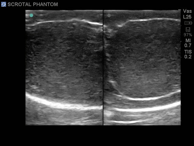
Blue Phantom ultrasound training model for scrotal ultrasound
Blue Phantom ultrasound training model for scrotal ultrasound
Blue Phantom's scrotal ultrasound training model offers users an excellent training platform for teaching and learning scrotal ultrasound examinations with their own ultrasound system. This extremely lifelike ultrasound phantom contains anatomically correct internal and external anatomy with both normal and pathological scrotal structures. Excellent for training in patient setup, image acquisition, interpretation, and the psychomotor skills required for scrotal ultrasound examinations – whenever you want, wherever you want, without risk to patients.
Gain competence in scrotal ultrasonography including patient setup and identification of normal structures and pathology
Blue Phantom's scrotal ultrasound training model contains the external anatomy of the male lower pelvis, penis, and scrotum. The contents of the scrotum are fully imageable with real-time 2D ultrasound and contain the left and right testes with epididymis (head, body, and tail) as well as pathological scenarios including intratesticular masses, an epididymal mass, and a hydrocele.
Ultrasound skills practiced include: use of ultrasound system settings, transducer positioning and movement, recognition of scrotal anatomy and pathology, and use of ultrasound calculation packages to measure cysts, masses, and other structures. The model accurately mimics the feel and imaging characteristics of a real scrotal ultrasound examination.
The model works well with all ultrasound systems with an appropriate scrotal transducer. Excellent for specialties such as urology, emergency medicine, fertility training programs, and medical educational facilities.
- Use your own ultrasound system for extremely lifelike scrotal ultrasound imaging
- Anatomically correct internal and external anatomy with both normal and pathological scrotal structures
- Excellent for training in patient setup, image acquisition, and interpretation
- Gain competence in scrotal ultrasonography including identification of normal structures and pathology
- Perform scrotal ultrasound examinations whenever and wherever you want – without risk to patients
- Contains normal testis and epididymis as well as contralateral testis and epididymis with pathology for comparison
- Contains external anatomy of the male lower pelvis, penis, and scrotum
- Fully imageable with real-time 2D ultrasound
- Left and right testes with epididymis as well as pathological scenarios including intratesticular masses, epididymal mass, and hydrocele
- Excellent sonographic imaging characteristics
- No replacement parts needed
- Tissue matches the acoustic properties of human tissue
- Works with all ultrasound systems
- Practice using ultrasound system settings
- High quality
- No special storage requirements
- Patented technology
- Made in the USA
- Includes soft storage/carry case
SKU BPS-801
Couldn't load pickup availability
- 1-3 day delivery
- Free shipping on purchases over DKK 3,000 excluding VAT
- Professional advice and guidance
Har du brug for hjælp? Kontakt vores kundeservice